MRI
Our Vision
To establish a distinguished research program that integrates basic development and clinical applications of MR imaging, spectroscopy, and MR-guided interventions for improved patient outcomes.
Our Mission
To develop and evaluate diagnostic, prognostic, and therapeutic MRI technologies including quantitative imaging that can be translated into clinical practice for improved patient outcomes and/or contribute to the knowledge of human physiology and diseases.
About Us
The MRI Research program in the Department of Radiology brings together a number of internationally recognized laboratories and investigators in the basic development and clinical applications of MR imaging, spectroscopy, and MR-guided interventions. The goals of the program are to carry out fundamental research that may lead either to the development of new techniques and interventions using MRI or to an improved understanding of the pathophysiology of disease for diagnosis and treatment. One of the important goals is to translate these advances rapidly into clinical practice, thereby improving the fundamental capabilities of diagnostic, prognostic, and theranostic imaging for improved patient care.
- Faculty & Staff
Faculty & Staff

Assistant Professor

Assistant Professor

Associate Professor
Research Lab
Professor

Assistant Professor
- Research Projects
Research Projects
Arterial Spin Labeled (ASL) MRI

Tissue perfusion is an important physiological parameter. Existing methods require administration of exogeneous contrast agent to measure tissue perfusion. Madhuranthakam Lab is developing ASL MRI methods that can measure tissue perfusion without the need for exogenous contrast agent. They are currently evaluating ASL-MRI in a variety of pathologies including brain tumors, kidney cancer, lung imaging and placenta imaging. Pedrosa Lab is also evaluating ASL as part of the multiparametric MRI for characterization of kidney cancer.
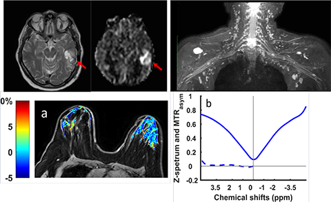
Chemical Exchange Saturation Transfer

CEST is a novel imaging method that allows the measurement of molecular characterization and alterations that are currently inaccessible by conventional MRI. Vinogradov Lab is developing and evaluating CEST in breast cancer, kidney cancer, and in neurodegenerative diseases such as multiple sclerosis and Alzheimer's.
Fat/Water Separation and Fat Quantification

Fat appears bright on conventional MRI and often confounds the visualization and characterization of underlying anatomical structures and pathologies. Madhuranthakam Lab is developing advanced imaging methods to achieve uniform fat/water separation without increasing scan times, and applying towards whole-body imaging. Dr. Yokoo is optimizing and evaluating fat quantification methods for accurate characterization of fatty liver disease.
High Temporal and Spatial Resolution (4D) MRI

High quality images with high spatial and temporal resolution that captures the transfer of exogenous contrast bolus provides quantitative parameters for accurate characterization of tumors. Dr. Guo and Pedrosa Lab are optimizing and evaluating 4D MRI techniques in liver cancer.
Inhomogeneous Magnetization Transfer (ihMT)

ihMT is a novel imaging method that allows detection and visualization of molecular characteristics specific to myelin. Vinogradov Lab is developing and evaluating ihMT in brain, spinal cord, and optic chiasm.
Multiparametric Imaging of Kidney Cancer

Pedrosa Lab focuses on evaluating novel MRI techniques for identification of vascular MRI measures that correlate to aggressiveness and molecular measures of angiogenesis, and the measures of tumor perfusion as an indicator of initial and acquired resistance to antiangiogenic therapies in metastatic disease.
Radiogenomics

Dr. Udayakumar is applying radiogenomics to decipher molecular and genomic alterations that are correlative to quantitative MRI features using pre-clinical models. She is extending these to human models in kidney cancer, in collaboration with Pedrosa Lab, and in breast cancer in collaboration with Vinogradov Lab.
Statistical Analysis

Dr. Xi supports various projects in the MR research program and in the Department of Radiology for statistical analysis. He is also developing pipelines for radiomics analysis and advanced statistical models to use radiological features as prognostic and predictive biomarkers.
Susceptibility Artifact Correction

Over 95% of orthodontic appliances are stainless steel that introduce magnetic susceptibility artifacts interfering with clinical brain MRI examinations. Wang Lab is developing methods including field correction devices using permanent magnets that can be used to correct for these artifacts without the need to remove the orthodontic appliances.
- Associated Cores
Associated Cores
DASPA
The Data Storage, Processing & Analysis core provides informatics support to researchers in a variety of areas.
Research PACS
- iPACS provides a HIPAA-compliant PACS system for archiving clinical and preclinical research imaging studies
- The installed and supported iPACS system is a web-based, secure, project-oriented resource available to Faculty researchers in Radiology and their collaborators
- Also has the capability of performing customized de-identification of images acquired in clinical trials to preserve patient confidentiality
ANSIR
The Advanced NeuroScience Imaging Research (ANSIR) lab dataset processing provides:
- Fully automated analysis of neuro MRI data including structural analysis using SPM and Freesurfer, task and resting state fMRI processing, Diffusion Tensor image processing, automated FLAIR white matter lesion segmentation, Arterial Spin Label MRI processing, and quantitative susceptibility mapping.
- Project-specific XNAT database storage allowing retrieval and queries of imaging and metadata
- Clinical alerts without a formal report can be provided for incidental findings
- Additional custom project-specific services may be available following consultation and based on resource availability
IM4T
The UTSW Radiology Department's Imaging Metrics for Trials (IM4T) group provides diagnostic imaging interpretation of cancer treatment response evaluation to internal and external researchers involved in cancer research studies that require response evaluation using Response Evaluation Criteria in Solid Tumors (RECIST) and its variants.
Magnetoencephalography (MEG)
- State-of-the-art technology mapping brain function
- Most advanced MEG technology currently available, and the only MEG scanner in Dallas
- Peripheral equipment available for time-locked stimuli and responses (ear buds, button pad, accelerometers, etc.)
- Used to study various neurological disorders and injuries including dementia, autism, concussion, and many others
Magnetic Resonance Imaging (MRI)
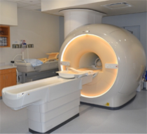
The Magnetic Resonance (MR) core is established to facilitate research and development within the Department of Radiology and collaborating departments in the field of MR imaging (MRI) and MR spectroscopy (MRS). The MR core consists of a whole-body human scanner (Philips Ingenia 3T MR) and a small animal Desktop scanner (Aspect 1T MR). Philips Ingenia includes dual-transmit and digital architecture for signal reception. This scanner enables the development and evaluation of new MRI/MRS techniques for improved diagnosis and understanding of the pathophysiology of disease. Aspect 1T scanner enables sequential imaging of small animals (e.g. mice and rats), without sacrificing them, for longitudinal monitoring of disease progression and/or therapy response. Being cited next to the small animal PET/CT and SPECT/CT scanners, this allows superposition of images for multimodality analysis.
Click here to explore more about the MR Core.
Translational Molecular Imaging Core (TMIC)
- Cyclotron and radiochemistry facility approved for CGMP production of PET radiopharmaceuticals for human use. Capable of producing 6 radioisotopes and >30 radiotracers in addition to the FDA-approved tracers
- A regulatory office in the Department of Radiology facilitates Investigational New Drug (IND) and Abbreviated New Drug Applications (ANDA) approval of radiotracers.
- Radiochemistry and nuclear medicine experts to advise investigators on the development and implementation of imaging protocols in a range of disease models (e.g., cancer, diabetes, metabolism, cardiotoxicity, neurodegenerative diseases, etc.)
- Pre-clinical imaging including a Siemens Inveon PET/CT scanner for small animal imaging
- State-of-the-art human imaging in the NE2 building including a GE Discovery IQ five ring PET/CT scanner and a Siemens 3T Biograph hybrid PET/MR scanner, both located in close proximity to our cyclotron facility
- Publications