PAAICI
About Us
The Program in Applied Artificial Intelligence in Clinical Imaging (PAAICI) in the Department of Radiology reflects the broad range of work occurring in the department in the areas of basic discovery, translation and clinical applications of artificial intelligence and machine learning in patient care.
PAAICI provides and coordinates the infrastructure to help faculty and trainees realize their goals in the development of new approaches in artificial intelligence A(I) and machine learning (ML) and move these new ideas into clinical practice. In addition, it provides venues to facilitate collaboration and shared knowledge amongst investigators.
Our Vision and Mission
The Department of Radiology has made significant investments to support the goals and vision of the PAAICI. These include:
- Leveraging the computational infrastructure of the BioHPC high performance computing center at UT Southwestern. This allows investigators to begin work without making a major, up-front investment in computing hardware or software.
- I want BioHPC (requires login)
- Implementing a research PACS or image archive system (iPACS) to facilitate the extraction of images from clinical PACS systems for de-identification, transformation and annotation. This research system is housed on BioHPC to facilitate processing.
- iPACS
- iPACS request form (requires login)
- Developing a Radiology Data Warehouse which includes millions of Radiology reports and associated metadata which can be used to identify cohorts of patients for AI/ML projects.
- Radiology Data Warehouse
- I want Data request form (requires login)
- Creating standardized paths for initiating relationships with external AI/ML companies to facilitate implementation and testing of algorithms developed externally. These also provide platforms for integration with clinical systems to make introduction of investigator developed algorithms into clinical workflow feasible.
- I want an agreement request form (requires login)
- Fostering collaboration with other artificial intelligence and machine learning initiatives at UT Southwestern. This includes developments in clinical and radiologist decision support through interaction with the Center for Clinical Informatics at UT Southwestern and the IT departments at tour partner hospitals.
- Faculty & Staff
Faculty & Staff
Professor
Vice Chair, Informatics
Professor
Research Lab
Professor
Director, Magnetoencephalography Program
Research Lab - Research Projects
Research Projects and Areas of Interest
- Neuroimaging - ANSIR
- Organ segmentation - Prostate, Liver, Kidney, Placenta
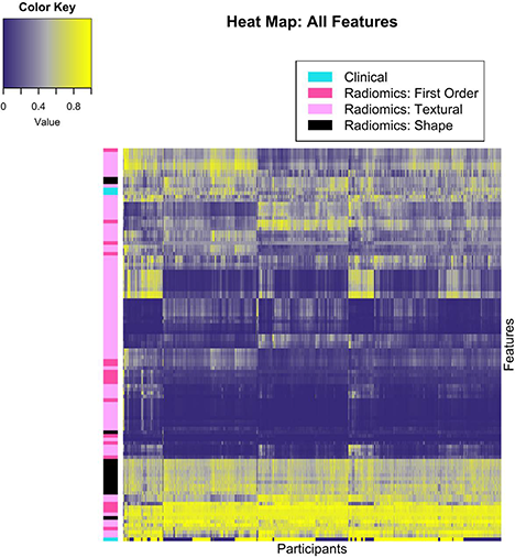
- Cancer genomics and radiomics - Kidney
- Cardiovascular disease detection and prognosis - Heart
- Opportunistic screening - Cardiovascular, Liver
- Workflow - ICH, PE
- Image Reconstruction - Hyper resolution
- Associated Cores
Associated Cores
DASPA
The Data Storage, Processing & Analysis core provides informatics support to researchers in a variety of areas.
Research PACS
- iPACS provides a HIPAA-compliant PACS system for archiving clinical and preclinical research imaging studies
- The installed and supported iPACS system is a web-based, secure, project-oriented resource available to Faculty researchers in Radiology and their collaborators
- Also has the capability of performing customized de-identification of images acquired in clinical trials to preserve patient confidentiality
ANSIR
The Advanced NeuroScience Imaging Research (ANSIR) lab dataset processing provides:
- Fully automated analysis of neuro MRI data including structural analysis using SPM and Freesurfer, task and resting state fMRI processing, Diffusion Tensor image processing, automated FLAIR white matter lesion segmentation, Arterial Spin Label MRI processing, and quantitative susceptibility mapping.
- Project-specific XNAT database storage allowing retrieval and queries of imaging and metadata
- Clinical alerts without a formal report can be provided for incidental findings
- Additional custom project-specific services may be available following consultation and based on resource availability
IM4T
The UTSW Radiology Department's Imaging Metrics for Trials (IM4T) group provides diagnostic imaging interpretation of cancer treatment response evaluation to internal and external researchers involved in cancer research studies that require response evaluation using Response Evaluation Criteria in Solid Tumors (RECIST) and its variants.
Magnetoencephalography (MEG)
- State-of-the-art technology mapping brain function
- Most advanced MEG technology currently available, and the only MEG scanner in Dallas
- Peripheral equipment available for time-locked stimuli and responses (ear buds, button pad, accelerometers, etc.)
- Used to study various neurological disorders and injuries including dementia, autism, concussion, and many others
Magnetic Resonance Imaging (MRI)
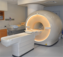
The Magnetic Resonance (MR) core is established to facilitate research and development within the Department of Radiology and collaborating departments in the field of MR imaging (MRI) and MR spectroscopy (MRS). The MR core consists of a whole-body human scanner (Philips Ingenia 3T MR) and a small animal Desktop scanner (Aspect 1T MR). Philips Ingenia includes dual-transmit and digital architecture for signal reception. This scanner enables the development and evaluation of new MRI/MRS techniques for improved diagnosis and understanding of the pathophysiology of disease. Aspect 1T scanner enables sequential imaging of small animals (e.g. mice and rats), without sacrificing them, for longitudinal monitoring of disease progression and/or therapy response. Being cited next to the small animal PET/CT and SPECT/CT scanners, this allows superposition of images for multimodality analysis.
Click here to explore more about the MR Core.
Small Animal Imaging Resource (SAIR)
- SAIR is an institutional facility that promotes and facilitates small animal imaging related to models of human disease with state-of-the-art equipment including depth resolved or planar optical imaging (fluorescence (FLI), bioluminescence (BLI) and chemiluminescence (CLI)), MRI, ultrasound, photoacoustic tomography, PET/CT, SPECT/CT and planar scintigraphy
- Infrastructure for animal handling (e.g., anesthesia, infusion, monitoring vital signs)
- Experienced investigators and technical staff capable of undertaking imaging and assisting in data interpretation are associated with the Resource and provide consultation on experimental planning, analysis and validation, and data archiving.
- Expertise in pulse programming and implementation for novel MRI experiments, design and acquisition or building of MR coils, choice of reporter molecules and /or genes, radiolabeling procedures and synthesis of ligands.
- Currently administered jointly by the Advanced Imaging Research Center (AIRC) and the Department of Radiology
Translational Molecular Imaging Core (TMIC)
- Cyclotron and radiochemistry facility approved for CGMP production of PET radiopharmaceuticals for human use. Capable of producing 6 radioisotopes and >30 radiotracers in addition to the FDA-approved tracers
- A regulatory office in the Department of Radiology facilitates Investigational New Drug (IND) and Abbreviated New Drug Applications (ANDA) approval of radiotracers.
- Radiochemistry and nuclear medicine experts to advise investigators on the development and implementation of imaging protocols in a range of disease models (e.g., cancer, diabetes, metabolism, cardiotoxicity, neurodegenerative diseases, etc.)
- Pre-clinical imaging including a Siemens Inveon PET/CT scanner for small animal imaging
- State-of-the-art human imaging in the NE2 building including a GE Discovery IQ five ring PET/CT scanner and a Siemens 3T Biograph hybrid PET/MR scanner, both located in close proximity to our cyclotron facility
- Publications
Publications