Quantitative Light Microscopy
Provide access to high-end optical microscopes to enable cancer-focused research.
C. Kern Wildenthal Research Building (NL)
NL 5 - North Campus
6000 Harry Hines Blvd.
Dallas, TX 75390
The Quantitative Light Microscopy Shared Resource (QLMSR) provides affordable access to a variety of state-of-the-art optical microscopes to support the cancer-focused research of Simmons members. The team consults with new and current users, provides customized training, and advises on sample preparation, data acquisition, and data analysis.
Services
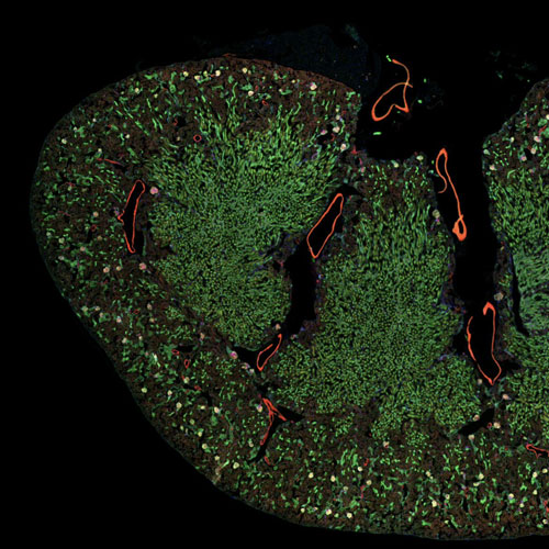
QLMSR offers a wide range of imaging modalities like wide-field microscopy, polarized light microscopy, state-of-the-art spinning disk and laser scanning confocal microscopy, and Total Internal Reflection Fluorescence microscopy.
QLMSR is the primary source for high-quality super-resolution imaging on the UT Southwestern campus and offers various modalities including AiryScan, SoRa, STORM, PALM, SIM, as well as access to a dedicated STED system. It provides instrumentation and expertise that permit multiday, time-lapse imaging of live samples and two photon microscopy, including second harmonic generation for visualizing collagen, muscle, and bone in unstained samples. It is the only source at UTSW for fluorescence correlation spectroscopy (FCS) in cells and solutions.
Users have 24/7 access to all imaging platforms after adequate customized training, as well as three powerful workstations and software for image processing and data analysis. The staff can work with users to develop customized image acquisition and analysis pipelines for, among others, colocalization, single particle tracking, and object counting and characterization.
An additional service made possible by QLMSR, in conjunction with the UTSW Electron Microscopy Core Facility, is correlative light and electron microscopy. Finally, in collaboration with other groups and core facilities, QLMSR provides access to cutting edge microscope technology for Simmons Cancer Center members that are not directly offered by the Shared Resource. QLMSR receives significant institutional support as well as support from the Simmons Cancer Center.
Technology and Equipment
The QLMSR maintains 13 microscopes, three image analysis workstations with software for image processing and analysis, and network-accessible servers for data transfer and temporary backup.
Instruments
- Zeiss LSM780 Upright scanning confocal/multiphoton
- Zeiss LSM780 Inverted scanning confocal/multiphoton
- Zeiss LSM880 Inverted scanning confocal/multiphoton and FCS
- Zeiss LSM880 Inverted scanning confocal with Airyscan
- Zeiss LSM980 Inverted scanning confocal with Airyscan2, AI sample finder, FCS and IR laser
- Nikon CSU-W1 SoRa spinning disk confocal with super resolution and FRAP modules
- Nikon CSU-W1 spinning disk confocal with 2 cameras
- Abberior Facility Line STED microscope
- GE OMX SR super-resolution microscope for 3D SIM, PALM/STORM and ring-TIRF
- Nikon Ti wide-field epifluorescence
- Zeiss AxioObserver epifluorescence
- Zeiss Axioscop histology
- Zeiss SteREO Discovery V12
- 3 powerful workstations for image analysis
- mesoSPIM for cleared specimens (isotropic ~5 µm resolution through very large volumes)
Fees
Simmons Cancer Center members and staff receive free training and a 25% subsidy for usage fees.
For additional information, including signing up for services, please visit the QLM Lab Site.
Leadership and Contact