
SPECT/CT
These are some sample SPECT/CT images from the Small Animal Imaging Resource (UT-SAIR).
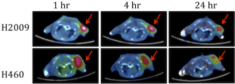
Lung cancer imaging: SPECT images showing different uptake dynamics of a 99mTc tagged with a DTPA tracer (i.p.) by H2009 vs. H460 tumors.
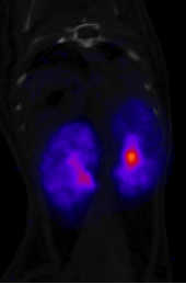
Renal imaging (SPECT/CT) using 99mTc-DMSA: When administered, 99mTc-DMSA intravenously apparently binds to plasma proteins in the blood and collects in the renal cortex. This allows measurement of relative left and right renal (kidney) function and is a very sensitive test to indicate the presence of renal scars or active infection in the kidney (pyelonephritis).
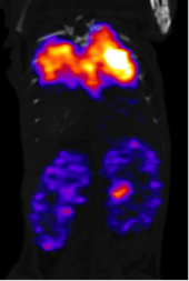
Renal imaging (SPECT/CT) using 99mTc-MAG3: The MAG3 clearance is highly correlated with the effective renal plasma flow (ERPF), and can be used as an independent measure of renal function. After intravenous administration, about 40-50% of the MAG3 in the blood is extracted by the proximal tubules with each pass through the kidneys; the proximal tubules then secrete the MAG3 into the tubular lumen. Note: MAA was also injected.
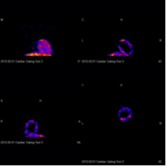
Cardiac gating is used to reduce motion artifacts generated by the pumping heart. ECG signals guide the SPECT acquisition so that the resulting images show the heart as it contracts.
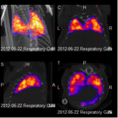
Respiratory gating: The respiration signals are monitored continuously and are used to guide SPECT acquisition the same way as cardiac gating.