Molecular Imaging and Probe Development
Molecular imaging techniques directly or indirectly monitor and record the spatiotemporal distribution of molecular or cellular processes for biochemical, biologic, diagnostic, or therapeutic applications. Mostly these methods utilize contrast agents (also called probes or tracers) that either specifically bind to the molecular target of interest, or resemble a metabolite detectable by in vivo imaging itself.
Inside AIRC, our primary interests focus on four main areas:
- Biologically responsive lanthanide-based MRI agents
- Stable isotope tracers of metabolism
- Novel hyperpolarized 13C and 15N MRI probes
- Genetically encodable probes for functional MRI – hemogenetic fMRI
Biologically Responsive Lanthanide-Based MRI Agents
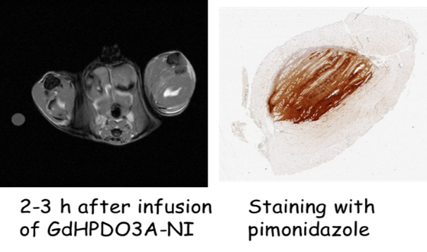
We are developing novel lanthanide complexes that report specific tissue biological information such as pH, oxygen levels, redox chemistry, enzyme activity, or metabolite concentrations. These agents include gadolinium based Zn2+ sensors for monitoring beta-cell function in the pancreas in vivo and for detecting prostate cancer by MRI. In addition, europium, terbium, and ytterbium containing paramagnetic chemical exchange saturation transfer (paraCEST) reporter systems can perform molecular imaging of pH, hypoxia, or tissue redox state. The image at right shows a T1-weighted image of a mouse tumor after infusion of a Gd-based hypoxia sensor developed in the AIRC. The bright region within the tumor reflects accumulation of the agent only in hypoxic regions. This was verified by a common histological marker of hypoxia (AD Sherry and X Wang, unpublished data).
Contact PI: A. Dean Sherry, Zoltan Kovacs
Stable Isotope Tracers of Metabolism
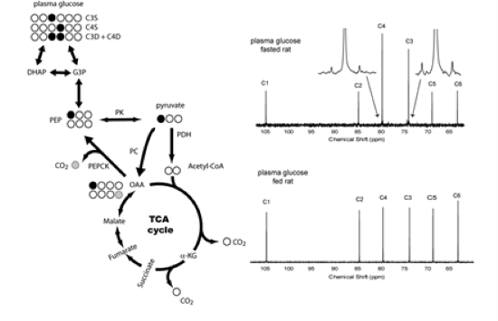
Metabolic substrates containing 2H or 13C labels in specific locations monitor relative flux through various competing pathways in vivo. These labeled molecules are either infused or given orally to animals or human subjects and metabolic information is obtained by performing conventional high-resolution NMR and/or mass spectrometry analysis of blood, urine, or tissue biopsy samples. The figure shows 13C NMR spectra of plasma glucose collected from a fasted rat (top chart at right) versus well-fed rat (bottom chart at right) after injection of [1-13C]pyruvate. Excess enrichment of glucose C3 and C4 in the top spectrum verifies that gluconeogenesis was highly active in fasted animals and less active in fed animals (ES Jin, et al. NMR Biomed, 2016, 29, 466-474).
Contact PI: Eunsook Jin, Craig Malloy, Anke Henning
Novel Hyperpolarized 13C and 15N MRI Probes
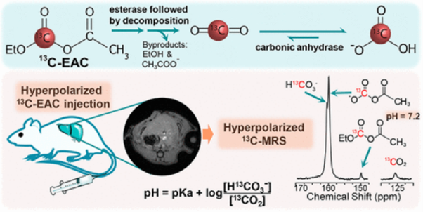
The emerging technology Dynamic Nuclear Polarization (DNP) can increase the sensitivity of NMR active nuclei by several orders of magnitude (50,000-fold or more), thereby allowing the real-time observation of biochemical processes in vivo. The chemistry group in the AIRC designs new 13C-enriched substrates which, after polarization by DNP, can be injected for imaging fundamental metabolic processes such as glycolysis, gluconeogenesis, pentose phosphate pathway activity, TCA cycle flux, or beta-oxidation. In addition to 13C-enriched probes, we also look into other NMR-active nuclei such as 15N and 89Y. These can be polarized and used as sensors of pH, redox, or zinc levels in tissue. The figure below illustrates the use of hyperpolarized 13C-enriched ethyl acetyl carbonate (13C-EAC), which hydrolyzes quickly once injected into animals to produce HP bicarbonate. The ratio of HP bicarbonate/HP CO2 provides a direct measure of tissue pH (Maptue, et al., ACS Sensors, 2018, 3, 2232-2236).
Contact PI: Zoltan Kovacs, A. Dean Sherry
Genetically Encodable Probes for Functional MRI – Hemogenetic fMRI
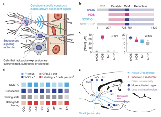
We developed a new functional MRI approach in which a genetically encoded probe converts intracellular calcium signals of targeted cells into hemodynamic changes detectable by MRI readouts. Thus, we named this technology hemogenetic fMRI.
The first generation of hemogenetic fMRI reporter is nitric oxide synthase for targeting image contrast (NOSTIC), a chimera of rat neural nitric oxide synthase (nNOS) and human inducible nitric oxide synthase (iNOS) proteins. The calcium sensitivity of nNOS can be activated by calcium influx into the cell membrane. NOSTIC inhibitor suppresses the activity of NOSTIC explicitly, but not that of endogenous nNOS, thus we used it to differentiate NOSTIC fMRI from endogenous fMRI background signals. We are working on other neurovascular coupling pathways for the next generation of hemogenetic fMRI probes. (N. Li*, S. Ghosh*, et al., Nat Neurosci. 2022, 25(3):390-398)
Contact PI: Nan Li