Protein plays dual role in causing, preventing sepsis
UTSW-led research could lead to new treatments for deadly complications from systemic infection
DALLAS – March 17, 2025 – A protein called angiopoietin-2 (ANGPT2) can both inhibit and encourage blood vessel changes critical for sepsis, a leading cause of hospital deaths worldwide, a new study led by UT Southwestern Medical Center researchers shows. Their findings, published in The Journal of Clinical Investigation, provide insight into how those changes occur and could lead to new ways to predict, monitor, and treat this condition.
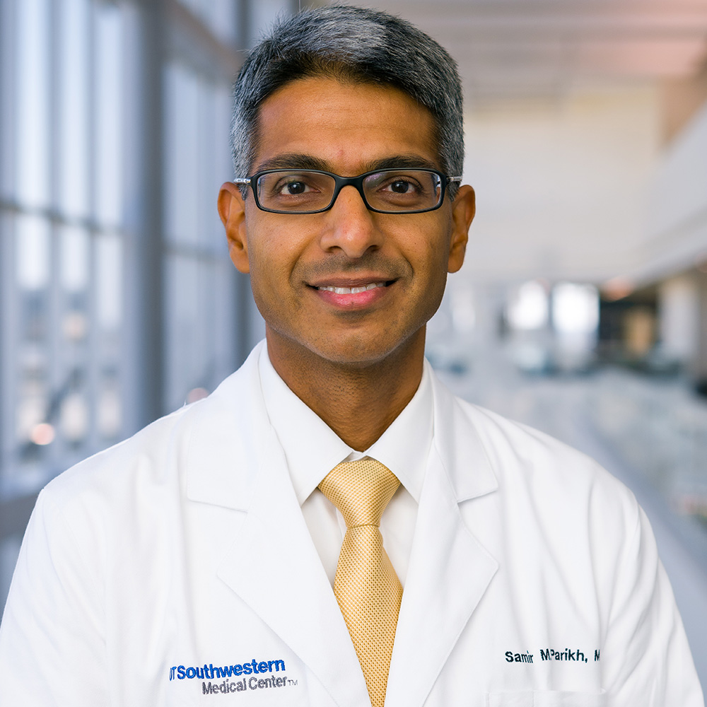
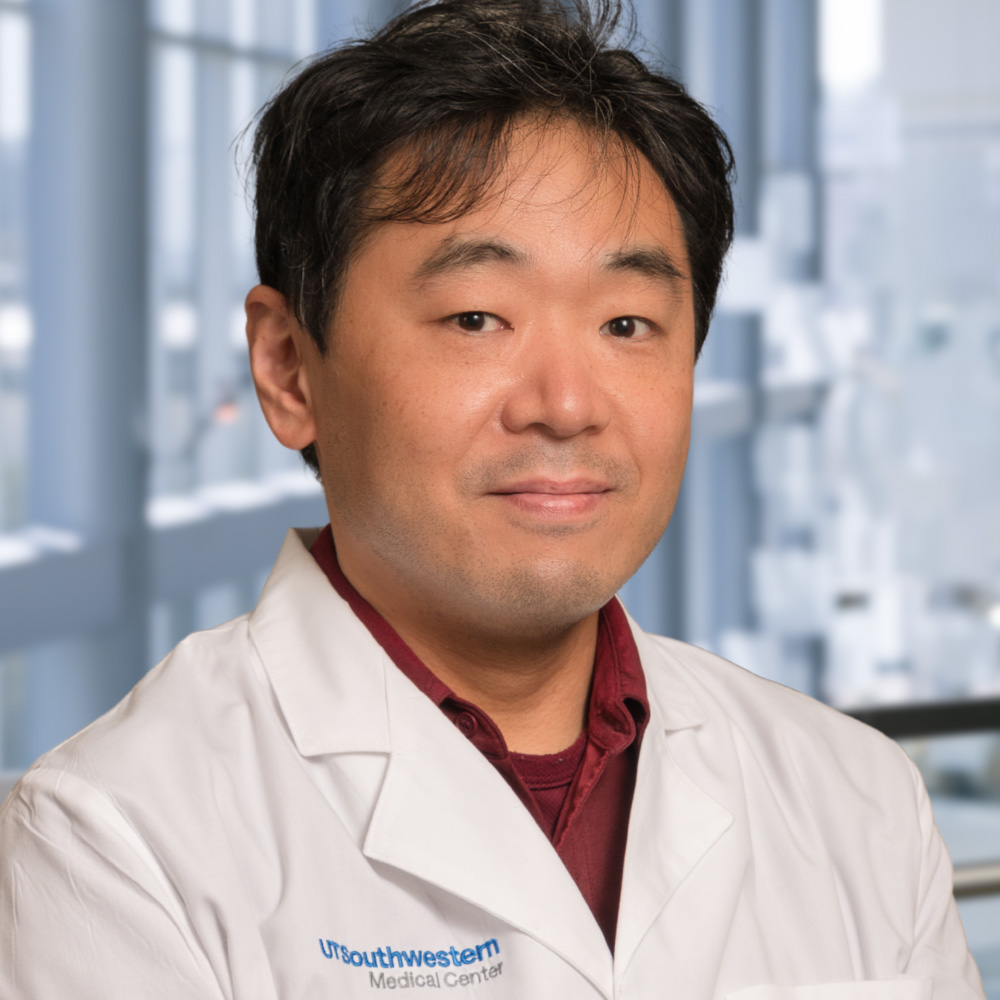
“People who die of sepsis are most often affected by multi-organ shutdown. Our research suggests that focusing on how small blood vessels fail under the stress of severe infection may open new opportunities to improve the health and resilience not just of blood vessels but also of major organs and therefore the entire body,” said Samir Parikh, M.D., Professor of Internal Medicine and Chief of the Division of Nephrology at UT Southwestern. Dr. Parikh co-led the study with Takashi Suzuki, Ph.D., Instructor of Internal Medicine in the Division of Nephrology.
In healthy people, Dr. Parikh explained, the endothelial cells that line blood vessels have anti-inflammatory properties to prevent blood from clotting and circulating immune cells from crossing over into adjacent tissues. This state is mediated by a receptor on endothelial cells called Tie2. Under normal conditions, ANGPT2 prompts Tie2 to produce an anti-inflammatory response.
However, these conditions are reversed during sepsis, an overwhelming response to systemic infection that can cause organ failure and sometimes death. Septic blood vessels promote clots and become leaky, swelling organs with water and releasing inflammatory cells from the blood. Previous research has shown that patients with sepsis overproduce ANGPT2, which in this state appears to block Tie2’s normal anti-inflammatory action. How ANGPT2 plays this dual role has been unknown.
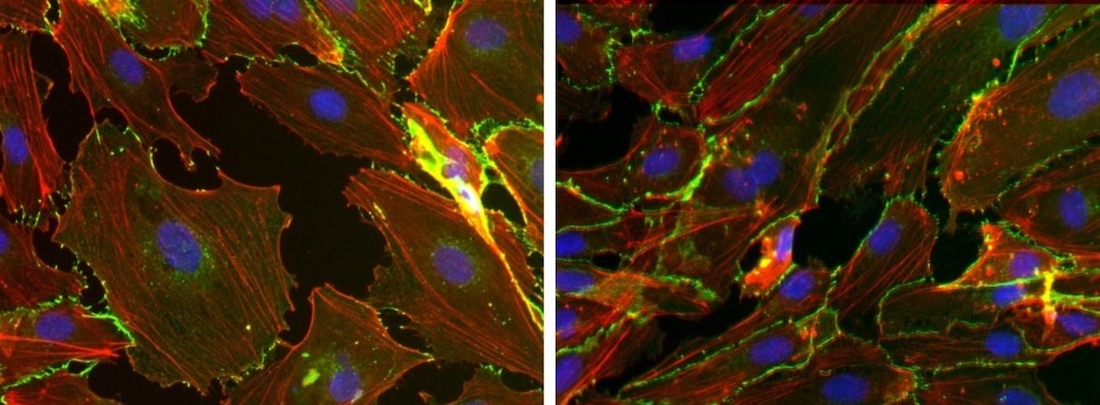
To answer this question, Drs. Suzuki and Parikh and their colleagues treated mouse immune cells with lipopolysaccharide, a molecule produced by some infectious bacteria. They then transferred the liquid these cells lived in, called culture media, to petri dishes where endothelial cells grew. The endothelial cells quickly switched from anti-inflammatory to inflammatory behavior, suggesting that something in this liquid had changed ANGPT2 from a protective role to a damaging one.
A closer look showed that the inflammatory ANGPT2 was no longer an intact molecule but had instead broken up into smaller pieces. Although intact ANGPT2 prevents blood vessel inflammation, the ANGPT2 fragments encouraged it.
Further experiments revealed that an enzyme called cathepsin K (CATK), produced by the immune cells, had chopped up ANGPT2 released by the inflamed endothelial cells. When the researchers applied an experimental drug that blocked CATK to these immune cells, the inflammatory response was no longer activated in endothelial cells. Mouse models of sepsis that were treated with the same drug retained significantly better lung and kidney function than those that didn’t receive the drug.
When the researchers examined the blood of patients who were septic, they found ANGPT2 fragments, suggesting that the same mechanism was at play. Consequently, Dr. Suzuki said, blood levels of ANGPT2 fragments and CATK could eventually be used to monitor the severity of patients’ conditions or their risk of sepsis. Still to be determined is whether human sepsis patients would benefit from the CATK-blocking drug, he said.
“If future clinical trials to use this drug to treat sepsis are successful, we would have a first-of-its-kind opportunity to save lives in the intensive care unit,” Dr. Suzuki said.
Drs. Parikh and Suzuki are listed as inventors on a patent filed by UT Southwestern related to this research in hopes of spurring the development of new diagnostic and therapeutic approaches in sepsis and other inflammatory conditions.
Other UTSW researchers who contributed to the study include Amanda J. Clark, M.D., Assistant Professor of Pediatrics; Vamsidhara Vemireddy, M.D., Research Program Manager; Erik Loyde, B.S., Research Associate; Valerie Etzrodt, M.D., Ph.D., Postdoctoral Researcher; and Marie Christelle Saade, M.D., Clinical Fellow.
Dr. Parikh holds the Ruth W. and Milton P. Levy, Sr. Chair in Molecular Nephrology and the Robert Tucker Hayes Distinguished Chair in Nephrology, in Honor of Dr. Floyd C. Rector, Jr.
This study was funded by an Outstanding Investigator Award from the National Heart, Lung, and Blood Institute (R35-HL139424) and grants from the National Institutes of Health (R01-DK095072 and R01-AG027002).
About UT Southwestern Medical Center
UT Southwestern, one of the nation’s premier academic medical centers, integrates pioneering biomedical research with exceptional clinical care and education. The institution’s faculty members have received six Nobel Prizes and include 25 members of the National Academy of Sciences, 23 members of the National Academy of Medicine, and 14 Howard Hughes Medical Institute Investigators. The full-time faculty of more than 3,200 is responsible for groundbreaking medical advances and is committed to translating science-driven research quickly to new clinical treatments. UT Southwestern physicians provide care in more than 80 specialties to more than 120,000 hospitalized patients, more than 360,000 emergency room cases, and oversee nearly 5 million outpatient visits a year.