Researchers make molecular connection between blindness, dementia
UTSW findings could lead to new ways to improve brain health in people with sensory impairments
DALLAS – Feb. 24, 2025 – Researchers at UT Southwestern Medical Center have linked blindness in animal models to a brain-wide cellular stress response that’s a common risk factor for dementia. Their findings, published in Nature Communications, could help explain the connection between vision or hearing loss and dementia.

“Our study provides mechanistic insight into how sensory impairment could be a potential risk factor for dementia and also emphasizes the importance of restoring or maintaining sensory systems to minimize complications,” said Helmut Krämer, Ph.D., Professor of Neuroscience and Cell Biology at UT Southwestern. Dr. Krämer co-led the study with Katherine Wert, Ph.D., Assistant Professor of Ophthalmology and Molecular Biology, and Shashank Shekhar, Ph.D., a postdoctoral fellow in the Krämer Lab.
Over the past decade, researchers have discovered that people with vision or hearing loss are significantly more likely to develop dementia. Studies have estimated that hearing loss in midlife contributes to 8.2% of dementia cases worldwide, and vision impairment is associated with 1.8% of U.S. dementia cases. However, the biological mechanism behind this link has been unknown.
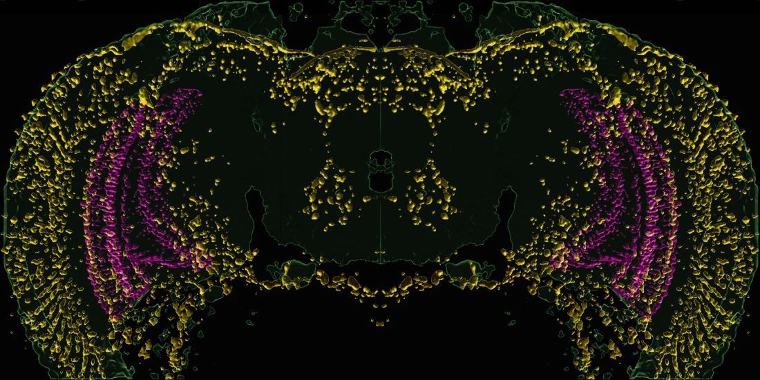
While studying molecular signaling in fruit flies, Drs. Krämer and Shekhar and their colleagues made an accidental discovery: The brains of fruit flies genetically altered to be blind had a widespread integrated stress response (ISR).
ISR protects cells throughout the body from conditions including starvation, viral infection, and oxygen deprivation by restricting cells’ protein production and turning on genes that fix cellular damage, Dr. Krämer explained. If ISR remains in effect for extended periods, it can kill cells through apoptosis – a mechanism to eliminate damaged cells that are unlikely to recover and threaten neighboring cells. Consequently, ISR is associated with neurodegenerative disorders, including Alzheimer’s disease and other forms of dementia.
Although the researchers saw ISR-propagating proteins known as ATF4 and XRP1 throughout the brains of the blind flies, they were in an unlikely place. Rather than being in the nucleus of affected cells, where they typically influence genes involved in ISR, ATF4 and XRP1 were found in molecular condensates. These droplets within a cell remain separate from its other contents, much like oil in water. When researchers in the Wert Lab looked at the brains of mice carrying genetic mutations that induced blindness, they saw a similar phenomenon.
The condensates bore a close resemblance to stress granules, a similar droplet that forms inside nerve cells under stress. When the researchers used a drug known to dissolve these stress granules, it also dissolved the condensates that bound ATF4 and XRP1, suggesting the condensates and stress granules might share a common purpose.
It’s not yet clear how such condensates might spur dementia if they develop in the brains of patients with blindness or other sensory impairments, Dr. Krämer said. One idea is that preventing proteins that propagate ISR from entering the cell nucleus might initially be protective, preventing long-term ISR that could cause apoptosis. However, the condensates could restrict ISR-associated proteins from responding to other neural complications, such as the overproduction of proteins associated with Alzheimer’s disease, making cells more vulnerable to damage.
Future studies will assess whether dissolving the molecular condensates could have a positive or negative effect on neurodegeneration, Dr. Wert said.
Drs. Krämer and Wert are members of the Peter O’Donnell Jr. Brain Institute and the Hamon Center for Regenerative Science and Medicine. Charles Tracy, M.S., Senior Research Associate at UTSW, also contributed to the study.
This research was funded by grants from the National Institutes of Health (R01EY10199, R01EY033184, R01AI155426, R21EY034597, and P30EY030413) and the Van Sickle Family Foundation.
About UT Southwestern Medical Center
UT Southwestern, one of the nation’s premier academic medical centers, integrates pioneering biomedical research with exceptional clinical care and education. The institution’s faculty members have received six Nobel Prizes and include 25 members of the National Academy of Sciences, 23 members of the National Academy of Medicine, and 14 Howard Hughes Medical Institute Investigators. The full-time faculty of more than 3,200 is responsible for groundbreaking medical advances and is committed to translating science-driven research quickly to new clinical treatments. UT Southwestern physicians provide care in more than 80 specialties to more than 120,000 hospitalized patients, more than 360,000 emergency room cases, and oversee nearly 5 million outpatient visits a year.