Study reveals cellular recycling process key to human health
UTSW-led research team uses cryo-EM to show how protein receptors return to a cell’s surface
DALLAS – Feb. 03, 2025 – A team led by UT Southwestern Medical Center researchers has identified a key mechanism responsible for endosomal recycling in cells, a process critical to human health. Their findings, published in Nature Communications, answer a fundamental question in cell biology and could lead to therapies for conditions including neurological disorders and cancer.
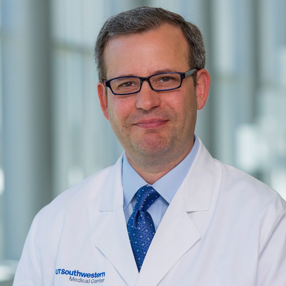
“Our study is a significant advance in understanding how proteins are recycled from endosomes back to the plasma (cell) membrane,” said Emre Turer, M.D., Ph.D., Associate Professor of Internal Medicine and Immunology at UT Southwestern. Endosomes play a vital role in the sorting and delivery of proteins and lipids within cells.
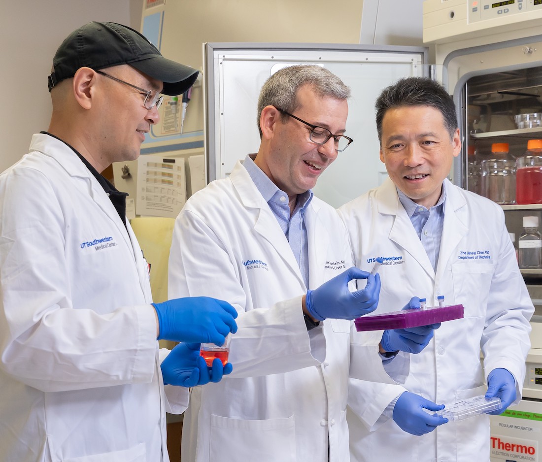
Dr. Turer co-led the study with Ezra Burstein, M.D., Ph.D., Professor of Internal Medicine and Chief of the Division of Digestive and Liver Diseases, and Baoyu “Stone” Chen, Ph.D., Associate Professor at Iowa State University.
Cell surfaces teem with many types of proteins; up to 10% of all proteins in the human body are at the cell surface. A significant portion of these are receptors – proteins that bind to molecules outside the cell and internalize them, returning to the cell surface to repeat the cycle. Known as endocytic recycling, this process is essential to cellular tasks such as nutrient uptake, intercellular communication, adhesion, and migration. Scientists have tied its disruption to health problems such as craniofacial and heart birth defects and certain types of immunodeficiencies.
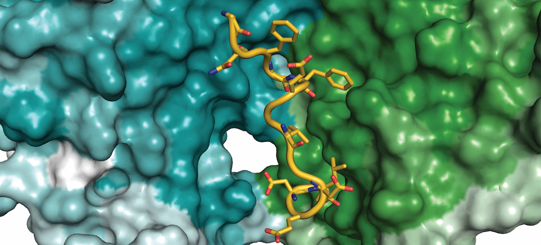
Although the internalizing of receptors has been well studied, Dr. Turer said, significantly less was known about how receptors make their way back to the cell surface. Previous research indicated that a protein called sorting nexin 17 (SNX17) and a protein complex called Retriever are pivotal to this recycling process, but how they work together has been unclear.
To answer this question, the researchers started by altering parts of SNX17 to better understand which section of the protein binds to Retriever. These experiments suggested that one end of SNX17 – called its C-terminal tail – attaches to another part of this protein. But when receptors pull in other molecules, these “cargoes” bind to SNX17 and displace the C-terminal tail, leaving it available to bind with Retriever.
The researchers then used cryogenic electron microscopy (cryo-EM) to view SNX17 bound to Retriever at a nearly atomic level, with results interpreted by cryo-EM expert Zhe “James” Chen, Ph.D., Professor of Biophysics and Co-Director of the Structural Biology Lab at UT Southwestern. This structure confirmed that SNX17’s C-terminal tail binds to Retriever, revealing a binding pocket on Retriever where this interaction happens.
When the researchers mutated Retriever’s binding pocket, SNX17 could no longer bind to it in a solution. Scientists identified 40 cell surface proteins that were significantly reduced in cells carrying a mutant version of Retriever with an altered binding pocket. Further research showed Retriever’s binding pocket is evolutionarily ancient, with an identical structure shared by organisms including humans, fruit flies, and amoebas.
Intriguingly, additional experiments identified 14 other proteins that appear to bind to this same pocket in Retriever because they carry a section akin to SNX17’s C-terminal tail. A search in a database of other protein structures suggests several other proteins may also interact with Retriever, including proteins from pathogens.
“Learning the roles of these other binding partners for Retriever could keep us busy for years to come,” said Dr. Burstein, who holds the Berta M. and Dr. Cecil O. Patterson Chair in Gastroenterology.
Other UTSW researchers who contributed to this study include first author Amika Singla, Ph.D., Assistant Professor of Internal Medicine; Xiaochen Bai, Ph.D., Associate Professor of Biophysics and Cell Biology; Ho Yee Joyce Fung, Ph.D., Assistant Professor of Biophysics; Yan Han, Ph.D., Assistant Professor of Biophysics; Andrew Lemoff, Ph.D., Associate Professor of Biochemistry; Ran Song, Ph.D., Senior Research Scientist; and Esther Banarer, Student Intern.
Drs. Turer and Burstein are members of the Harold C. Simmons Comprehensive Cancer Center.
This study was funded by grants from the National Institutes of Health (R35 GM128786, R01 DK107733, R01 DK133229, and R01 DK119360), The Welch Foundation (I-1944), and the National Science Foundation (CDF 2047640).
About UT Southwestern Medical Center
UT Southwestern, one of the nation’s premier academic medical centers, integrates pioneering biomedical research with exceptional clinical care and education. The institution’s faculty members have received six Nobel Prizes and include 25 members of the National Academy of Sciences, 24 members of the National Academy of Medicine, and 14 Howard Hughes Medical Institute Investigators. The full-time faculty of more than 3,200 is responsible for groundbreaking medical advances and is committed to translating science-driven research quickly to new clinical treatments. UT Southwestern physicians provide care in more than 80 specialties to more than 120,000 hospitalized patients, more than 360,000 emergency room cases, and oversee nearly 5 million outpatient visits a year.