UTSW study reveals how key protein affects neuron structure
Insight into function of torsinA furthers understanding of rare movement disorder called DYT1 dystonia
DALLAS – Sept. 20, 2024 – A protein called torsinA plays a key role in the early development of neurons, determining where nuclear pores are placed in the membrane that encloses the nucleus of nerve cells, a study led by UT Southwestern Medical Center researchers shows. Their findings, published in Nature Cell Biology, answer long-standing questions about torsinA’s function and could lead to treatments for a rare movement disorder called DYT1 dystonia, caused by a torsinA mutation.

“The function of torsinA has been elusive. This work demonstrates that torsinA is involved in the spatial organization of nuclear pore complexes and suggests dysregulation of this process during neurodevelopment could contribute to long-lasting neuronal deficits,” said Samuel Pappas, Ph.D., Assistant Professor in the Peter O’Donnell Jr. Brain Institute and of Neurology at UT Southwestern.
Dr. Pappas co-led the study with William Dauer, M.D., Director of the O’Donnell Brain Institute and Professor of Neurology and Neuroscience, and Sami Barmada, M.D., Ph.D., Associate Professor of Neurology at the University of Michigan. The study’s first author is Sumin Kim, Ph.D., a former graduate student at the University of Michigan who was co-mentored there by Drs. Pappas, Dauer, and Barmada.
DYT1 dystonia is marked by abnormal twisting and tremors in the arms and legs that arise in childhood. Epidemiological studies have estimated this inherited condition affects 54,000 to 80,000 people in the U.S. However, nearly three times this number carry the genetic mutation in torsinA that causes DYT1 dystonia. Why symptoms arise during this period, how mutations in torsinA cause it, and why so many carriers are unaffected are unknown. The function of torsinA is a critical question tying these mysteries together, Dr. Pappas said.
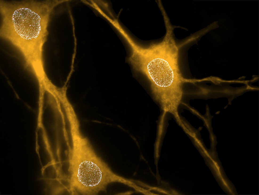
In earlier studies, he and his colleagues learned that deleting the gene that makes torsinA early in development in animal models, but not in adulthood, caused neurons to develop nuclear pore complexes (NPCs) – collections of proteins that form nuclear envelope pores – grouped in clusters, rather than distributed across the nuclear membrane. These small openings in the nuclear envelope allow proteins and genetic material to move between the cell nucleus and the liquid that fills cells, called cytoplasm.

To determine how torsinA might be involved in NPC distribution, Drs. Pappas, Dauer, Barmada, and their colleagues tracked the number and placement of these structures in neurons growing in petri dishes isolated from animals soon after birth. Within the first few days after isolation, these cells made significantly more NPCs, which correlated with maturing of neuronal circuits. Additional experiments in cultured neurons demonstrated that deleting torsinA from these cells didn’t change the number of NPCs, but it did change where they were placed in the nuclear envelope, causing the clustering the researchers had seen in the previous study.
To investigate the function of torsinA in live animals, the researchers developed a special mouse line in which one protein in the NPC was tagged with a fluorescent marker. They saw that the clusters were made of newly formed NPCs that were not distributed evenly across the nuclear membrane. Further research showed that neurons in which the gene for torsinA was deleted or replaced with the same mutated form carried by patients with DYT1 dystonia developed abnormal swollen areas in the nuclear envelope before NPCs arose during maturation. These “blebs” were located in the same places containing clusters of NPCs. Using a technique called super-resolution microscopy, the researchers showed that the resulting NPCs appeared to have a normal conformation, albeit with an abnormal, clumped distribution in the nuclear envelope.
Together, Dr. Pappas explained, these findings suggest torsinA controls where NPCs are ultimately placed during a critical period early in neuron development – a step that appears key to healthy neuronal function later in life and matches with the timeline of when DYT1 dystonia symptoms typically arise.
Further study could help scientists understand what differs between people who carry the disease-causing mutation and develop symptoms and those who never do. Eventually, he said, these findings might lead to therapies to treat this disease.
“Our prior work showed that torsinA impairment, as occurs in dystonia, plays an essential role in the formation of the central nervous system during a critical period in development,” Dr. Dauer said. “This new work identifies nuclear pore biogenesis as a torsinA-dependent process during that critical period, so it is exciting in establishing a mechanistic focus to overcome this defect.”
Other UTSW researchers who contributed to this study include Sarah Shahmoradian, Ph.D., Assistant Professor in the Center for Alzheimer’s and Neurodegenerative Diseases, of Biophysics, and in the O’Donnell Brain Institute; and Hung Tri Tran, Ph.D., Research Scientist.
Dr. Dauer holds the Lois C.A. and Darwin E. Smith Distinguished Chair in Neurological Mobility Research.
This study was funded by grants from the National Institutes of Health (R01 NS110853, R01 DK118480, R01 NS077730, NIH R01 NS097542, R01 NS113943, R56 NS128110, GM129347, T32 GM007315, U24 NS120055, 1S10OD021784, and R01 GM082949), the National Science Foundation (MCB1552439 and NSF2014862-UTA20-000890), and a University of Michigan Rackham Predoctoral Fellowship.
About UT Southwestern Medical Center
UT Southwestern, one of the nation’s premier academic medical centers, integrates pioneering biomedical research with exceptional clinical care and education. The institution’s faculty members have received six Nobel Prizes and include 25 members of the National Academy of Sciences, 22 members of the National Academy of Medicine, and 14 Howard Hughes Medical Institute Investigators. The full-time faculty of more than 3,200 is responsible for groundbreaking medical advances and is committed to translating science-driven research quickly to new clinical treatments. UT Southwestern physicians provide care in more than 80 specialties to more than 120,000 hospitalized patients, more than 360,000 emergency room cases, and oversee nearly 5 million outpatient visits a year.