Pathway tied to cancer-driving genome alterations identified
Understanding how chromosome shattering occurs could lead to strategies to overcome therapy resistance, UTSW study shows
DALLAS – Sept. 09, 2024 – Cancer cells appear to hijack a genetic pathway involved in DNA repair to drive malignancy and overcome treatment, a study led by UT Southwestern Medical Center researchers shows. Their findings, published in Cell, explain how chromosomes in some tumors undergo massive rearrangements and could lead to new strategies to avoid cancer drug resistance.
“Our research answers a key mechanistic question in cancer biology by identifying a source of chromothripsis, a mutational process driven by chromosome shattering. Chromothripsis allows cancer cells to rapidly evolve their genomes by extensively rearranging individual chromosomes, leading to multiple genetic alterations within a relatively short period of time,” said study leader Peter Ly, Ph.D., Assistant Professor of Pathology and Cell Biology at UT Southwestern and a member of the Cellular Networks in Cancer Research Program in the Harold C. Simmons Comprehensive Cancer Center.
Chromosome rearrangements caused by chromothripsis occur in 30%-40% of all cancers and are commonly found in aggressive tumors such as sarcomas, glioblastomas, and pancreatic cancer.
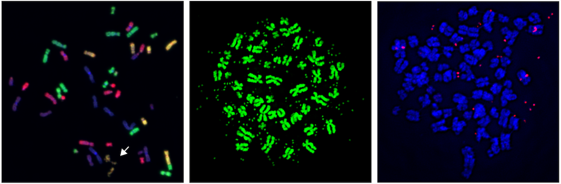
Chromothripsis can occur when mitosis, the process of cell division, goes awry. Cells can mistakenly sort whole chromosomes or chromosome arms outside of the nucleus and into abnormal packages called micronuclei. Previous work by the Ly Lab demonstrated that these chromosomes in micronuclei eventually shatter into many pieces and become reassembled in the incorrect order. How these chromosomes shatter has been unknown.
To answer this question, Dr. Ly, along with Justin Engel, B.S., a graduate student researcher in the Cancer Biology Ph.D. Program, used CRISPR, a gene-editing tool, to inactivate genes in cells with micronuclei to find those that might play key roles in chromosome shattering. Their hunt narrowed to a set of genes involved in the Fanconi anemia pathway – a DNA repair mechanism mutated in an eponymous germline condition marked by severe anemia, bone marrow failure, cancer predisposition, and other congenital defects. When the researchers inactivated these genes, the chromosomes didn’t shatter.
Further research showed that unlike the chromosomes in the nucleus, those in micronuclei fail to properly replicate, spurring the Fanconi anemia pathway into action. As part of this DNA repair process, an enzyme complex then chops these chromosomes into pieces, causing the chromosome shattering characteristic of chromothripsis.
“When these pieces are stitched back together in the incorrect order,” Dr. Ly explained, “they result in chromosome rearrangements that often inactivate genes meant to protect against cancer development. In addition, some pieces develop into a circular DNA structure called extrachromosomal DNA, or ecDNA, that gets amplified to a large number of copies, a process that can promote drug resistance.”
To extend their findings into a clinically relevant setting, Dr. Ly and his colleagues inactivated the Fanconi anemia pathway in melanoma tumor cells treated with targeted therapy. Although these cancer cells typically acquire resistance over time to these drugs, cells lacking the Fanconi anemia pathway didn’t undergo chromothripsis and failed to develop drug resistance.
“These findings could eventually lead to new strategies incorporating Fanconi anemia pathway inhibition with other drugs to combat the emergence of therapy-resistant cancer cells, which represents a significant problem for cancer patients,” Dr. Ly said.
Other UTSW researchers who contributed to this study are Qing Hu, Ph.D., Senior Research Associate, and Kidist Woldehawariat, B.S., Research Technician.
This study was funded by grants from the National Institutes of Health (NIH) (R01CA289435, R35GM146610), the Cancer Prevention and Research Institute of Texas (CPRIT) (RR180050), The Welch Foundation (I-2071-20240404), and a National Cancer Institute Cancer Center Support Grant (P30CA142543). Mr. Engel was supported by the CPRIT Cancer Intervention and Prevention Discoveries Program Training Grant (RP210041) and the NIH Ruth L. Kirschstein National Research Service Award Predoctoral Fellowship from the National Cancer Institute (F31CA295091).
About UT Southwestern Medical Center
UT Southwestern, one of the nation’s premier academic medical centers, integrates pioneering biomedical research with exceptional clinical care and education. The institution’s faculty members have received six Nobel Prizes and include 25 members of the National Academy of Sciences, 22 members of the National Academy of Medicine, and 14 Howard Hughes Medical Institute Investigators. The full-time faculty of more than 3,200 is responsible for groundbreaking medical advances and is committed to translating science-driven research quickly to new clinical treatments. UT Southwestern physicians provide care in more than 80 specialties to more than 120,000 hospitalized patients, more than 360,000 emergency room cases, and oversee nearly 5 million outpatient visits a year.