

Researchers unlock key step in cell fusion process
Understanding protein assembly during cell-cell fusion could ultimately aid in development of new therapies for various diseases
UT Southwestern researchers defined mechanisms by which cells create invasive protrusions to facilitate their fusion. These cellular protrusions are primarily generated by a protein called actin when it forms polymers, or filaments, that push the plasma membrane outward. The investigators pieced together a comprehensive picture involving a crew of proteins that makes branched actin filaments, organizes the branches into bundles, stabilizes the bundles, and ultimately makes invasive protrusions.
Understanding how invasive protrusions are created in cell-cell fusion helps researchers understand cancer metastasis and immune response and could ultimately lead to therapeutic approaches for a range of diseases, including congenital myopathies, cancer, and immune disorders.
“This study solves a decades-old problem in actin assembly: how branched actin filaments are organized into mechanically stiff protrusions,” said study leader Elizabeth Chen, Ph.D., Professor of Molecular Biology and Cell Biology, whose work focuses on cell-cell fusion.
Cell-cell fusion is indispensable for skeletal muscle development and regeneration. During embryonic development, individual muscle cells called myoblasts fuse to form multinucleated muscle fibers or myofibers. Myofibers are elongated cells that contain all the nuclei from a group of fused myoblasts. Myofiber formation allows many myoblasts to be controlled by nerves as a single unit. Myoblast fusion is also required to repair wounds or exercise-induced muscle injuries.

Back in 2010, Dr. Chen and her team discovered that myoblast fusion is an asymmetrical process that involves a myoblast projecting invasive protrusions into its fusion partner at the cell-cell interface, which they named the “fusogenic synapse.” The invasive protrusions used by myoblasts are similar to those used by cancer cells to penetrate surrounding tissues during their spread to distant organs and by white blood cells to traverse blood vessels during an immune response.
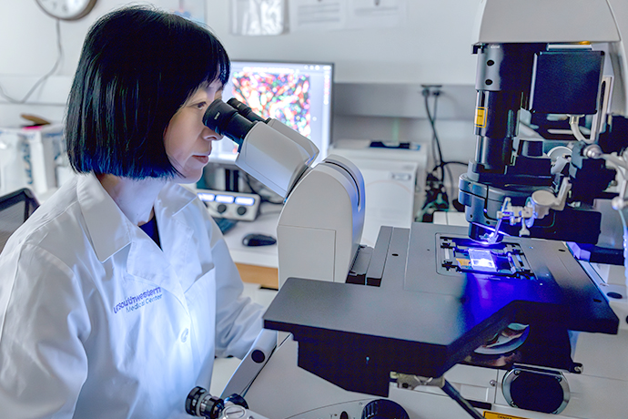
In the current study published in Nature Cell Biology, Dr. Chen and her team used live cell imaging, super resolution and transmission electron microscopy, mouse genetics, and other techniques to monitor actin assembly during invasive protrusion formation. They looked at which proteins are enriched at different locations along these protrusions and how they coordinate their actions during myoblast fusion.
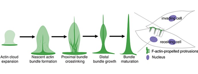
They found that two proteins, N-WASP and WAVE, which are known to activate a protein complex called Arp2/3 to generate branched actin filaments, are enriched in the shafts and tips of invasive protrusions. In the absence of these proteins, invasive protrusions are no longer generated by a cell. This is intriguing, Dr. Chen said, because “prior to this work, cellular protrusions, such as filopodia, were thought to be made from linear actin filaments. How branched actin filaments are organized to make invasive protrusions remained a mystery.”
The research team found that to make an invasive protrusion, the Arp2/3 complex first generates an “actin cloud” consisting of floppy actin filaments that branch and re-branch. Then a protein called dynamin enters the actin cloud to create order out of the disordered branched actin network. This is due to dynamin’s unique ability to form helical structures, each of which can recruit up to 16 actin filaments and organize them into a loose parallel bundle, according to Dr. Chen’s and her team’s earlier study. Once dynamin forms a full helical structure, it rapidly breaks apart and releases individual dynamin proteins back into the cytosol, leaving behind a loose actin bundle.
In the current study, the Chen Lab discovered that to tighten up the loosely aligned actin filaments, cells use an additional actin-crosslinking protein, the N-WASP-interacting protein WIP, to lock the actin filaments together into a mechanically stiff bundle. Thus, to build invasive protrusions from branched actin filaments, a cell needs to orchestrate the actions of four different proteins – two catalyzing actin filament formation and another two organizing the filaments into bundles – in a spatially and temporally coordinated manner.
“It would not be surprising that cancer cells and white blood cells use a similar mechanism to generate their invasive structures,” said Dr. Chen, who is also a member of the Hamon Center for Regenerative Science and Medicine and the Harold C. Simmons Comprehensive Cancer Center.
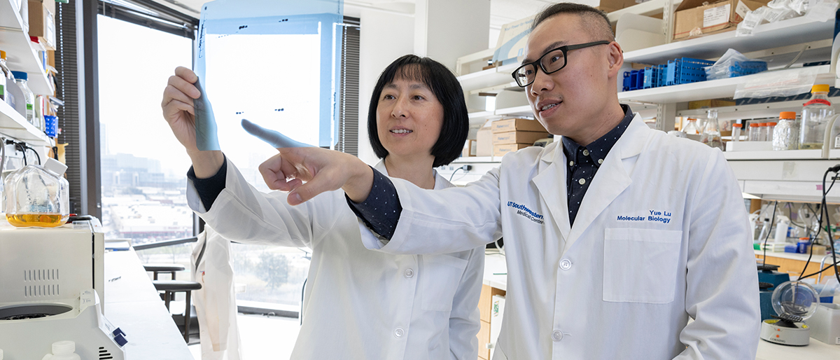
Contributors to this research from the Department of Molecular Biology include Yue Lu, Ph.D., Instructor; Benjamin Ravaux, Ph.D., and Zhou Luo, Ph.D., Assistant Instructors; Pratima Pandey, M.S., Chief Electron Microscopy Technician; Tezin Walji, a former graduate student researcher; and Ruihui Zhang, Ph.D., a former postdoctoral researcher. Other UTSW investigators are Bing Li, Ph.D., Research Scientist in Physiology; Chuanli Zhou, Ph.D., Senior Research Associate in Immunology; and Duojia Pan, Ph.D., Chair of Physiology and a Howard Hughes Medical Institute Investigator.
This work was supported by National Institutes of Health grants and a UT Southwestern Hamon Center for Regenerative Science and Medicine fellowship.
Endowed Title
Dr. Pan holds the Fouad A. and Val Imm Bashour Distinguished Chair in Physiology.