

Structural integrity
A world-renowned structural biologist and an equally esteemed scientist specializing in biochemical function combine their strengths to build lasting insights – and overcome challenges.
Structural biologist Dr. Xiao-chen Bai, Assistant Professor of Biophysics and Cell Biology, specializes in cryo-electron microscopy (cryo-EM). His interests include studying tyrosine kinase receptors such as the insulin receptor (IR). Despite decades of study, the activation mechanism for that receptor is largely unknown. Dr. Hongtao Yu, Adjunct Professor of Pharmacology, works to understand the function of the IR using biochemical and cell biology approaches. He published a Cell study in 2016 that established a major IR regulatory mechanism linking guardians of chromosome stability to nutrient metabolism.
Soon after Dr. Bai’s arrival at UTSW in 2017, he and Dr. Yu decided that investigating the structure of the human IR in the insulin-bound active state would benefit from their complementary approaches. As their study was in progress, Dr. Giovanna Scapin and colleagues at Merck & Co. published in Nature the low-resolution cryo-EM structure of a truncated IR and reported insulin binding at two locations.
Knowing how insulin binds to the receptor has implications for improving our understanding of insulin’s function. The UTSW researchers forged on, publishing in eLife their own work with the intact IR that showed insulin binding at up to four sites, including two never before identified. Their subsequent experience – though not uncommon – is seldom discussed with the candor with which they are willing to share with In Pursuit readers.
Why did you decide to study the structure of the IR bound to insulin?
Dr. Bai: The insulin receptor belongs to a family of tyrosine kinase receptors and plays essential roles in glucose metabolism and cell growth. Dysregulation of IR signaling is linked to diabetes, cancer, and other diseases. Since insulin was discovered in the 1920s, researchers have been working to understand its signaling pathway.
Surprisingly, despite nearly 100 years of study, how insulin activates the IR is still not fully understood. We think this is mainly due to the complexity of the way insulin binds to its receptor and also the challenge of solving the structure of transmembrane proteins. The activation of the IR requires multiple insulin molecules cooperatively binding to different sites. Therefore, to reveal the exact IR activation mechanism, it is necessary to know the detailed structure of the IR in its fully functional state. We set out to get the clearest, most complete structure of the receptor. We wanted to see the full-length receptor actively bound to insulin and to understand how the binding of multiple insulin molecules to distinct sites affects the activation of the receptor’s tyrosine kinase domain, which is located inside the cell.
When the other study came out, did you consider abandoning your project?
Dr. Bai: Yes, we considered stopping our project, but we saw that the Nature study had investigated a truncated receptor that contained only the extracellular domain. It was solved only at low resolution by cryo-EM. Also, when we looked at their data carefully, it didn’t seem to match the mutagenesis, binding, and biochemical data that we expected based on other groups’ previous studies. Finally, we had set out to solve the structure of the entire receptor, and we were still very curious to see if that could be done.
Insulin is a small molecule and is actually very difficult to make. It’s also difficult to make and purify the entire functional insulin receptor. As a single-pass transmembrane tyrosine kinase receptor, it exists in three environments: the oxygen-rich extracellular environment, the lipid-filled transmembrane domain, and the reducing, gel-like environment inside the cell. To see all three domains clearly using cryo-EM requires studying the protein under conditions that keep all three parts happy. That meant the use of detergents and buffer conditions that mimic the three environments, which may be why the structure of the entire receptor had not been determined prior to our work.
Dr. Yu: Also, there was a discrepancy between earlier work predicting two binding areas with different affinities and the Nature study, which indicated two binding areas with similar affinities.
Dr. Bai: Once we decided to go ahead with our project, we expected to at least confirm what the other team had found, though at a higher resolution. This would allow us to build a precise model for the entire receptor complex and pinpoint the detailed conformational change that occurs upon receptor activation.
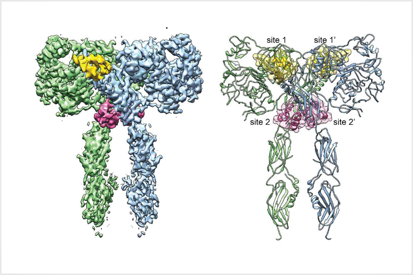
Then, as often happens in science, we found something unexpected. We found that up to four insulin molecules could bind to the receptor, two at the canonical site and two at a novel site. While the location of the canonical insulin binding site 1 of IR has been known for a long time, researchers have been looking for the second insulin binding site for many decades. It was very exciting when we realized that our data identified the location of site 2 on the IR for the first time. Our structure also provided some clues to explain why IR activation requires multiple insulin molecules bound at two different types of sites. Again, as often happens in science when a result is unexpected, some people in the field have had a difficult time believing our results.
Dr. Yu: We want to give special thanks to the co-lead authors of our study, Dr. Eunhee Choi of my lab and Dr. Emiko Uchikawa of the Bai lab, for their diligence in pursuing the IR project throughout these ups and downs.
Could you elaborate on the differences between the two studies’ findings?
Dr. Bai: The Scapin study in Nature, which reported a resolution of 4.3 angstroms (Å), indicated that the insulin receptor was a dimer with two equivalent insulin binding sites. In contrast, we observed at a higher resolution of 3.1Å that up to four insulin molecules can bind to the receptor. We referred to the binding sites identified in the Nature paper as site 1 and site 1′ and referred to the two new sites we identified as site 2 and site 2′. The two new sites had slightly lower affinity for insulin than do sites 1 and 1′. Ours was the first cryo-EM structure of the full-length insulin receptor with fully occupied insulin binding sites captured in the fully functional state.
For a long time, many people believed that only one insulin molecule is necessary and adequate to fully activate the insulin receptor. Based on our data, this is an incorrect belief. Our model shows that full activation of the insulin receptor requires three to four insulin molecules binding simultaneously at two distinct types of sites. After comparing our IR structure with those solved by others, we found that the binding of multiple insulin molecules to the two different types of sites in a cooperative manner promotes the structural transition of the IR from an inactive to an active state.
Dr. Yu: We did extensive mutagenesis and biochemical studies to confirm our results and then submitted our work for publication.
Dr. Bai: We had some trouble publishing this study. Our proposed model of insulin activation is striking and very different from all previous models, and thus was difficult for the initial reviewers to accept. Despite this opposition, we eventually got the study published in eLife.
What are the possible caveats or unanswered questions in this research?
Dr. Yu: One hesitancy is that we used very high concentrations of insulin to ensure maximum occupancy of the receptor bound to insulin in our samples. The question is whether such high concentrations of insulin ever occur under normal conditions. The Scapin study published in Nature used these same conditions in their work. In addition, both groups used liver cancer cells to study the IR, so it is unknown if other cell types might show different responses.
It appears from our work that you don’t need all four insulin molecules to turn on the receptor. It is still unknown and worth figuring out whether you need two, three, or four insulin molecules bound to the receptor for activation, and how different patterns of occupancy on the four sites we’ve identified might produce different outcomes in IR signaling pathways driving glucose metabolism under normal conditions or in Type 2 diabetes or those driving growth pathways in cancer.
Any regrets?
Dr. Yu: Our strongest motivation was to see more and see better – to visualize the entire insulin receptor and understand how insulin binding turns on the kinase domain inside the cell. Even though we solved the structure of the full-length receptor, we still couldn’t see the kinase portion of the protein and that’s something we eventually would like to do.
Future steps?
Dr. Bai: We are still working on it. Our final goal is to understand why insulin resistance happens, using a combination of structural and biochemical studies together with other approaches. Insulin receptor activation appears to be a multistep process. We want to know how different functional states and binding patterns can affect blood sugar metabolism.
Dr. Bai is a Cancer Prevention and Research Institute of Texas Scholar and a Virginia Murchison Linthicum Scholar in Medical Research.